Cardiovascular disease is still the leading cause of deaths in the UK, responsible for one in every three, even though the numbers have halved since the 1970s and 1980s. In 2010, about 80,000 deaths were caused by cardiovascular disease and 49,000 by strokes. Both conditions also cause a great deal of long-term damage and ill-health to people who survive.The NHS website states that coronary heart disease is the UK's biggest killer and causes around 82,000 deaths each year. About one in five men and one in eight women die from the disease. In the UK, there are an estimated 2.7 million people that live with the condition and a further 2 million affected by angina, the most common symptom of CHD. It generally affects more men than women by from the age of 50, the chances of developing CHD are similar for men and women. Angina is one of the main symptoms of CHD as well as heart attack and heart failure. Not everyone has the same symptoms and some people may not have any of these symptoms before CHD is diagnosed. It is sometimes called ischameic heart disease.
To explain heart disease and how it works, we should first consider the heart and how it works:


How the heart works:
- Each day, your heart beats about 100,000 times.
- It pumps about 23,000 litres (5,000 gallons) of blood around your body.
- This blood delivers oxygen and nutrients to all parts of your body, and carries away unwanted carbon dioxide and waste products.
- The heart is divided into two pumps which work together. Blood that is coming back from the organs and tissues of your body enters the right side of your heart which then pumps it to your lungs. Your lungs remove waste carbon dioxide from the blood and recharge it with oxygen. The oxygen-rich blood returning from your lungs enters the left side of your heart, which then pumps it to all parts of your body, including the heart muscle itself. This process ensures that there is always enough oxygen and nourishment for your body to work efficiently
- The heart comprises less than 0.5 percent of the total body weight and has three layers. The smooth, inside lining of the heart is called the endocardium. The middle layer of heart muscle is called the myocardium. It is surrounded by a fluid filled sac call the pericardium.
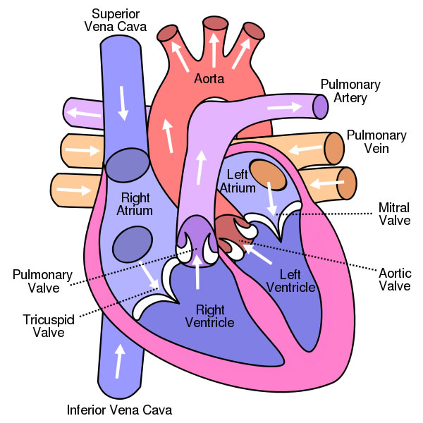
- It is divided into four chambers, the right atrium (RA), the right ventricle (RV), the left atrium (LA), and the left ventricle (LV). Each of the chambers has a sort of one-way valve at its exit, which prevents blood from flowing backwards. When each chamber contracts, the valve at its exist opens. Pressure on one side is higher than the other and that creates the flow forwards in one direction where it cannot go backwards. When it has finished contracting, the valve closes so that the blood does not flow backwards.
- The tricupside valve is at the exit of the right atrium, the pulmonary valve is at the exit of the right ventricle, the mitral valve is at the exit of the left atrium and the aortic valve is at the exit of the left ventricle.
- When the heart muscle contracts or beats, this action is called a systole. It pumps blood out of the heart. The heart contracts in two stages: in the first stage, the right and left atria contract at the same time, pumping blood to the right and left ventricles. Then the ventricles contract together to propel blood out of the heart. Then the heart muscle relaxes (called diastole) before the next heartbeat. This allows blood to fill up the heart again.
- The right and left sides of the heart have separate functions. The right side of the heart collects oxygen-poor blood from the body and pumps it to the lungs where it picks up oxygen and releases carbon dioxide. The left side of the heart then collects oxygen-rich blood from the lungs and pumps it to the body so that the cells throughout your body have the oxygen they need to function properly.
- Diagram of the chambers:

- All blood enters the right side of the heart through two veins: the superior vena cava and the inferior vena cava. The superior vena cava collects blood from the upper half of the body and the inferior vena cava collects blood from the lower half of the body. Blood leaves the two veins and enters the right atrium. When the right atrium contracts, the blood goes through the tricuspid valve into the right ventricle. When the RV contracts, blood is pumped through the pulmonary valve into the pulmonary artery and into the lungs where it picks up oxygen.
- Blood returning from the body is poor in oxygen as it has been oxygen has been diffusing into cells its been passing. So it needs to be full of oxygen before returning to the body. So the right side of the heart pumps blood to the lungs first, picking up oxygen, before going to the left side of the heart where it is returned to the body.
- It returns from the lungs to the heart via the pulmonary veins and goes into the left atrium. When the left atrium contracts, blood travels through the mitral valve and into the left ventricle. The left ventricle pumps blood from the aortic valve into the aorta. The aorta is the main artery of the body: it receives all the blood the heart has pumped out and distributes it to the rest of the body. The left ventricle has a thicker muscle because it must pump blood to the rest of the body against a higher pressures in general circulation.
Heart disease
- Coronary heart disease is the term that describes what happens when your heart's blood supply is blocked or interrupted by a build-up of fatty substances in the coronary arteries. Over time, the walls of your arteries can become furred up with fatty deposits. This process is known as atherosclerosis and the fatty deposits are called atheroma.
- This can be caused by lifestyle habits and other conditions, such as: smoking, high cholesterol, high blood pressure and diabetes
- When the heart muscle goes without sufficient oxygen, the muscle is said to be ischemic. If cell death occurs it is called infarction. Since a heart attack is cell death of heart muscle (myocardium), it is called a Myocardial Infarction (MI). The condition that causes CAD, angina and heart attacks is called atherosclerosis.
- Coronary arteries supply blood to the heart muscle. When a blockage occurs in one of these arteries, blood flow to the heart muscle is decreased. This will be realised during exertion as the heart muscle works harder and needs more oxygen-enriched blood than normal. By preventing the much needed increase in blood flow, the blockage deprives the heart muscle of oxygen thereby causing the heart muscle to hurt. This chest pain is called angina.
- Arteriosclerosis is a more general term for hardening of the arteries. Atherosclerosis is a type of arteriosclerosis that causes a buildup of fatty material along the inner lining of arteries. When a heart muscle goes without sufficient oxygen, the muscle is said to ischemic. If cell death occurs, it is called infarction. A heart attack is cell death of heart muscle and is aclled a myocardial infarction.
- So, in summary, if a blockage occurs in a coronary artery, it causes chest pain (angina), if the blockage is complete, it can cause a heart attack (myocardial infarction), if a blockage occurs in one of the arteries near the brain, a stroke can occur and if a blockage occurs in a leg artery, it causes peripheral vascular disease and can cause pain while walking. This is called intermittent claudication.
Hope this has been informative, thanks for reading
No comments:
Post a Comment